Center for Electron Microscopy
Transmission electron microscopy provides an insight into the ultrastructure of the cell, meaning that cell membranes, cell organelles and, for example, intercellular contact structures can be visualized. Viruses or nanoparticles can also be made visible and antigens can be marked using immunogold labelling.
At the Center for Electron Microscopy, we are available to all researchers at the School of Veterinary Medicine and other institutions within and outside of the Freie Universität Berlin.
The Center for Electron Microscopy is affiliated with other electron microscopy facilities in Berlin under the Alliance Center Electron Microscopy (ACEM).
Equipment and Analysis Method
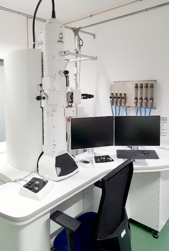
We have a powerful transmission electron microscope (JEM-1400-Flash, Jeol) and all the equipment for preparing, fixing and embedding your biological samples, as well as the ultramicrotomes required for the semi-thin and ultra-thin sectioning technique. With the help of various embedding and contrasting methods, we are able to work on a wide range of biological research questions.
Thanks to the experience of the well-coordinated team, transmission electron microscopy questions are dealt with professionally and, if required, co-users are supported with imaging and analysis.
We can develop concepts together with you, starting with:
- sampling procedure
- suitable fixative
- appropriate sample size
- structures to be examined
- optimal staining and contrasting protocols
- forms of embedding
- examination on the device and subsequent image processing
We are also able to compile series images and create tilt series for tomographic examinations.
If you are interested in the services we offer, please contact:
-
Contact persons: Claudia Kipar, Pascal-Kolja Bingül
-
Telephone: +49 (0)30 838 75947
Publications with Electron Microscopic Images from our Center for Electron Microscopy:
| Author, Titel, Journal | Reference & Hyperlink |
|---|---|
| Jevtić M, Löwa A, Nováčková A, Kováčik A, Kaessmeyer S, Erdmann G, Vávrová K, Hedtrich S. (2020) Impact of intercellular crosstalk between epidermal keratinocytes and dermal fibroblasts on skin homeostasis. Biochim Biophys Acta Mol Cell Res. 2020 Apr 14:118722. | doi.org/10.1016/j.bbamcr.2020.118722 |
| Kordowitzki P, Merle R, Hass K, Plendl J, Rieger J, Kaessmeyer S (2021) Influence of age and genetic background on bovine ovarian capillary blood supply, ovarian mitochondria, and telomere length. Cells, 10, 2661 | doi.org/10.3390/cells10102661 |
| Kanevche K., Burr D.J., Nürnberg D.J., et al. (2021) Infrared nanoscopy and tomography of intracellular structures. Commun Biol 4, 1341 | doi.org/10.1038/s42003-021-02876-7 |
| Zabihi F, Tu Z, Kaessmeyer S, Schumacher F, Rancan F, Kleuser B, Böttcher C, Ludwig K, Plendl J, Hedtrich S, Vogt A, Haag R. (2021) Efficient Skin Interactions of Graphene Derivatives: Challenge, Opportunity, or both? Pharmaceutics | In Revision |
| Jevtić M, Rieger J, Nováčková A, Kováčik A, Tan Z, Vávrová K, Hedtrich S, Kaessmeyer S (2022) Native Extracellular Matrix Significantly Improves the Differentiation and Maturation of Three-dimensional Skin Models. Cells | In Revision |
| Maneenooch K. (2022) PhD Dissertation FU Berlin Institut für Veterinär-Anatomie (Gast-PhD des Department of Anatomy, Faculty of Veterinary Medicine, Kasetsart University, Bangkok, Thailand) | From porcine skin samples in situ to three-dimensional human skin constructs in vitro. Studying skin with a focus on the 3R principles |
| Mechsner S. (2021) DFG Geschäftszeichen ME 3413/10-1 | Mechanismen der Neuroimmunomodulation im Rahmen der chronischen Inflammation bei Endometriose |
| Fisch S, Keilholz U, Tinhofer-Keilholz I (2021) "MSTARS"; Förderkennzeichen des Teilprojektes 1: 031L0220A Charité Comprehensive Cancer Center Multimodal clinical mass spectrometry to target treatment resistance | Multimodal clinical mass spectrometry to target treatment resistance |
| Alshamy Z, Richardson K, Harash G, Hünigen H, Röhe I, Hafez MH, Plendl Al Masri S (2019): Structure and age-dependent growth of the chicken liver together with liver fat quantification: a comparison between a dual-purpose and a broiler chicken line. PLoS One | doi: 10.1371/journal.pone.0226903 |
| Khiao In M, Richardson K, Loewa A, Hedtrich S, Kaessmeyer S, Plendl J (2019): Histological and functional comparisons of four anatomical regions of the porcine skin which human abdominal skin. Anat Histol Embryol, 48(3):207-217. | doi: 10.1111/ahe.12425 |
| Aguilera-Rojas M, Badewien-Rentzsch B, Plendl J, Kohn B, Einspanier R (2018): Exploration of serum- and cell culture-derived exosomes from dogs. BMC Vet Res. 2018 Jun 8;14(1):179 | doi: 10.1186/s12917-018-1509-x |
| Kaessmeyer S, Sehl J, Khiao In M, Hiebl B, Merle R, Jung F, Franke RP, Plendl J (2016): Organotypic soft-tissue co-cultures: Morphological changes in microvascular endothelial tubes after incubation with iodinated contrast media. Clin Hemorheol Microcirc 64(3):391-402 | doi: 10.3233/CH-168119 |
| Ibrahim MG, Chiantera V, Frangini S, Younes S, Köhler C, Taube ET, Plendl J, Mechsner S (2015): Ultramicro-trauma in the endometrial-myometrial junctional zone and pale cell migration in patients with adenomyosis. Fertil Steril 104: 1475-1483 | doi: 10.1016/j.fertnstert.2015.09.002 |